Modern dental technology allows us to make your dental visits more comfortable and efficient and allows us to provide excellent long-term outcomes. If you'd like to see all the ways we improve your dental experience, please check out our leading-edge technology.
Cephalometric (Ceph) X-Ray
A cephalometric or ceph x-ray is a unique diagnostic tool our dentist uses to capture a radiographic image of the side of your face. This type of x-ray is comfortable because it's taken outside the mouth (extraoral), and there's no need to bite down on any film.
We commonly use ceph x-rays when planning dental implant placement because they show the entire oral structure in one image. Viewing the ceph x-ray enables our dentist in Las Vegas to identify tooth fractures, jawbone injuries, and malocclusions (bite problems) to plan orthodontic treatment.
Panoramic X-Rays
A panoramic dental x-ray shows us your entire mouth in a single image, similar to taking a pano image on your phone. This x-ray provides a flat image of the jaw’s curved structure, including all your teeth, upper and lower jaws, temporomandibular joints (TMJs), and nasal and sinus cavities.
We commonly use panoramic x-rays to diagnose fractures, infections, impacted wisdom teeth, bone abnormalities, cysts, and tumors. These details are invaluable when planning dental implant placement, orthodontic treatment, and wisdom tooth extraction.
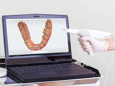
Digital Impressions
Taking dental impressions used to be a messy, uncomfortable process, but not anymore! Instead of the dreaded pink paste you may have had in the past, we use an intraoral scanner.
Getting a digital impression is easy because all you do is sit still while we move the scanner over your teeth. This leading-edge digital imaging technology instantly delivers a 3-D image of your teeth that our dentist views on a computer monitor.
There’s no sitting around with an uncomfortable tray filled with gooey paste stuck in your mouth and no waiting while the material sets. Digital impressions are more comfortable and provide a highly-detailed, accurate representation that is not possible with paste impressions.
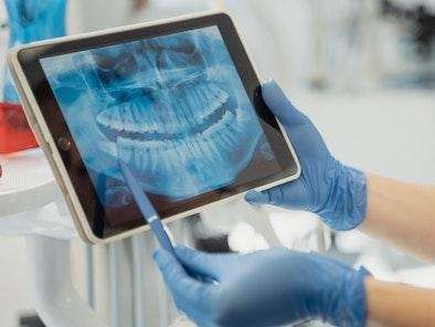
Digital X-Rays
Dental x-rays are an invaluable tool that allows us to see inside your teeth and under your gums, which isn’t possible with a visual examination. At Russell Dental, we care about your safety and use digital x-rays and sensors that expose you to a significantly lower radiation dose than film x-rays.
We share the digital images to show you what we see while explaining our diagnosis. By rotating, enlarging, or zooming in on the image, we can help you see the area in great detail, which helps you understand why we recommend treatment and how it will benefit your oral health.
Because we use digital records, we can easily and safely store your x-rays in your patient history, where our team members can access them easily. Digital x-rays also easier to share with your insurance company or specialists.

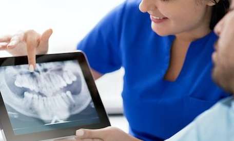
Intraoral Cameras
An intraoral camera is a technology we to show you real-time images of the inside of your mouth as we perform your exam. As we point the small digital camera at different parts of your mouth, you can see the images on your chairside monitor.
Our dentist uses the images to point out problems like cavities and broken or worn teeth, so you can see what he sees. Being told about a cavity is one thing, but seeing it helps you understand how treating it will benefit your oral health.
And after treatment, we’ll use the intraoral camera to show you the results! We can also store the images in your patient history, so they are always available as needed.
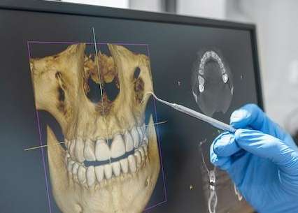
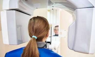
3-D Cone Beam Imaging (CBCT)
When we plan root canals, dental implant placement, and surgical tooth extractions, it's crucial to see the structures of your mouth in enhanced detail.
At Russell Dental, we use sophisticated 3-D cone beam digital imaging technology (CBT or CBCT). These scans provide three-dimensional images showing the position and location of sinuses, nerve pathways, and root canals, in addition to soft tissue and bone density.
When we see these features in detail, our dentist can plan and perform these procedures with an extremely high degree of precision and accuracy. Cone beam CT scans are invaluable when planning implant placement to determine the optimal location and position in your jaw to ensure positive long-term outcomes.

Digital Records
It’s important to us that your appointments run smoothly and efficiently. Using digital records, the team at Russell Dental creates a seamless workflow so everyone can easily access your patient history. There’s no sorting through mountains of files, and all your information is stored safely on our secure in-house network.